

File:Psoriatic arthritis fingers ar1934-1.gif
Size of this preview: 960 × 631 pixels. Other resolutions: 330 × 217 pixels | 1,004 × 660 pixels.
Original file (1,004 × 660 pixels, file size: 396 KB, MIME type: image/gif)
Captions
Captions
Add a one-line explanation of what this file represents
Summary
[edit]
| Description |
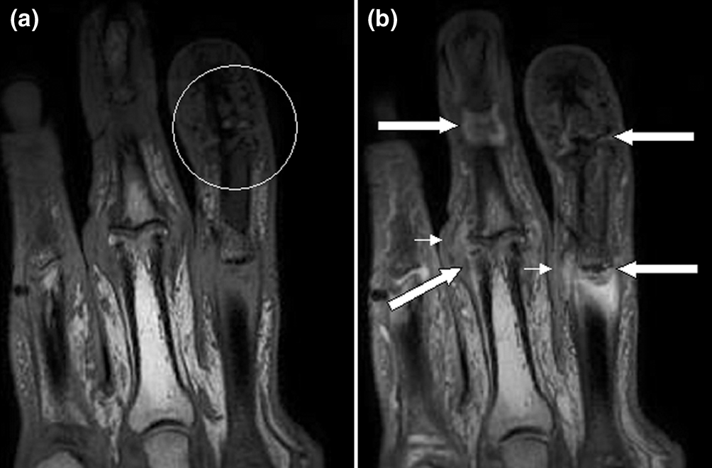
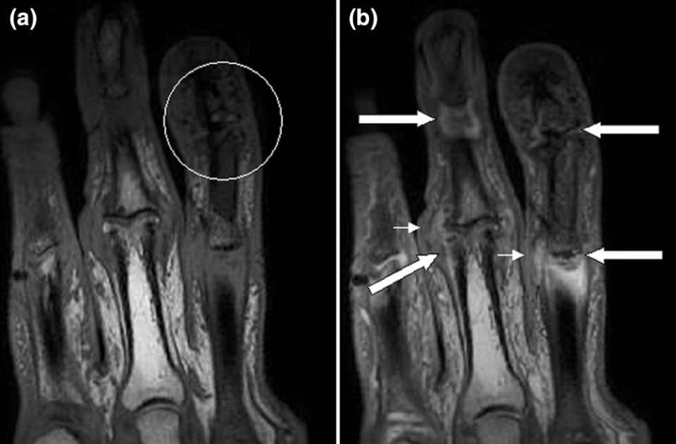
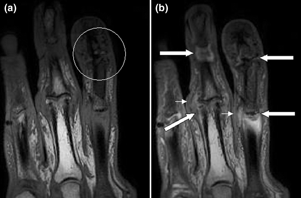
English: Magnetic resonance images of fingers: psoriatic arthritis. Shown are T1-weighted (a) precontrast and (b) postcontrast coronal magnetic resonance images of the fingers in a patient with psoriatic arthritis. Enhancement of the synovial membrane at the third and fourth proximal interphalangeal (PIP) and distal interphalangeal (DIP) joints is seen, indicating active synovitis (large arrows). There is joint space narrowing with bone proliferation at the third PIP joint and erosions are present at the fourth DIP joint (white circle). Extracapsular enhancement (small arrows) is seen medial to the third and fourth PIP joints, indicating probable enthesitis. Note that this particular slice does not allow optimal visualization of all of the mentioned pathologies. |
| Date | Published: 23 March 2006 |
| Source | Magnetic resonance imaging in psoriatic arthritis: a review of the literature. Arthritis Research & Therapy 2006, 8:207. doi:10.1186/ar1934 |
| Author | Fiona McQueen, Marissa Lassere and Mikkel Østergaard. |
Licensing
[edit]


This file is licensed under the Creative Commons Attribution 2.0 Generic license.
- You are free:
- to share – to copy, distribute and transmit the work
- to remix – to adapt the work
- Under the following conditions:
- attribution – You must give appropriate credit, provide a link to the license, and indicate if changes were made. You may do so in any reasonable manner, but not in any way that suggests the licensor endorses you or your use.
File history
Click on a date/time to view the file as it appeared at that time.
| Date/Time | Thumbnail | Dimensions | User | Comment | |
|---|---|---|---|---|---|
| current | 00:08, 1 June 2021 | | 1,004 × 660 (396 KB) | Jul059 (talk | contribs) | Lossless file size reduction |
| 10:49, 7 February 2009 | 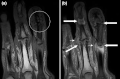 | 1,004 × 660 (422 KB) | Stevenfruitsmaak (talk | contribs) | {{Information |Description={{en|1=Magnetic resonance images of fingers: psoriatic arthritis. Shown are T1-weighted (a) precontrast and (b) postcontrast coronal magnetic resonance images of the fingers in a patient with psoriatic arthritis. Enhancement of |
You cannot overwrite this file.
File usage on Commons
There are no pages that use this file.
File usage on other wikis
The following other wikis use this file:
- Usage on ar.wikipedia.org
- Usage on bs.wikipedia.org
- Usage on en.wikipedia.org
- Usage on en.wikibooks.org
- Usage on hu.wikipedia.org
- Usage on outreach.wikimedia.org
- Usage on sr.wikipedia.org
- Usage on uk.wikipedia.org
- Usage on zh.wikipedia.org
